Nocturnal Arousal Mapping Reveals Sleep Disorder Signatures
THE PROTOHUMAN PERSPECTIVE#
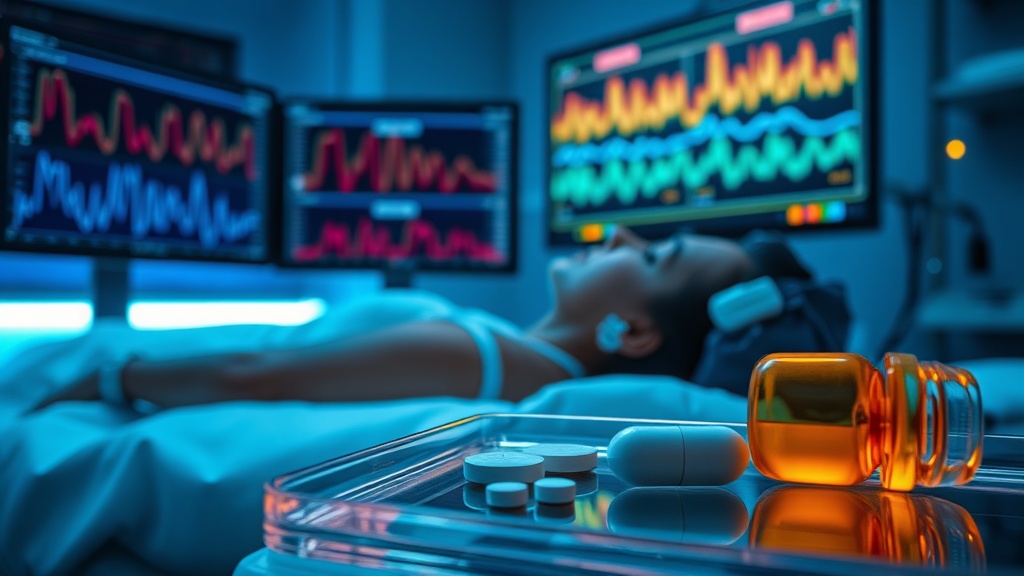
Sleep isn't a single state. It's a continuously shifting negotiation between arousal and descent — and we've been measuring it wrong for decades. The new mapping work from Biabani, Mendonça, Mutti et al. doesn't just add another sleep study to the pile. It reframes the entire question: instead of asking how much sleep someone gets, it asks how the brain's arousal machinery behaves minute-by-minute across the night, and how that machinery breaks down differently depending on whether you have fibromyalgia, narcolepsy, or a parasomnia.
For anyone serious about performance optimization, this matters. The nocturnal arousal trajectory may be a more sensitive biomarker of brain health than total sleep time or even sleep stage percentages. If we can fingerprint these trajectories with automated tools — and the wearable detection research is catching up fast — we move closer to genuinely personalized sleep interventions. Not "sleep more." Not "take magnesium." But targeted correction of the specific arousal coupling failure your brain exhibits.
THE SCIENCE#
What Is Nocturnal Arousal Coupling, Exactly?#
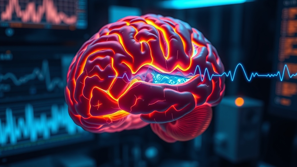
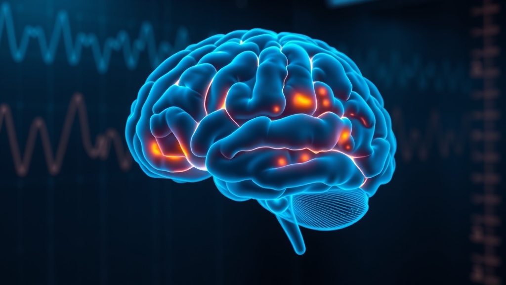
Nocturnal arousal coupling refers to the way the brain's intrinsic phasic activations — brief, transient bursts of cortical activity — interact with the homeostatic drive to deepen sleep as the night progresses[1]. In healthy sleepers, early-night NREM sleep is dominated by slow-wave activity (A1-type phasic events), which are thalamocortical in origin and reflect high homeostatic sleep pressure. As that pressure dissipates across the night, these slow-wave-dominant responses decay predictably.
This is the pattern the new study from Biabani et al. (2026) set out to map — and then compare across disorders[1].
The A-Phase Index: A New Lens on Sleep Microstructure#
The team used an automated metric they call the A-phase index (API), inspired by the cyclic alternating pattern (CAP) framework but designed to work without requiring B-phase identification. The API provides a 60-second rolling readout of phasic burden — essentially, how much arousal-related activity the brain is generating at any given point in the night[1].
— actually, I want to clarify something here, because the terminology can mislead. "Arousal" in this context doesn't mean waking up. It refers to brief, often subcortical activations that punctuate sleep without producing conscious awareness. They're part of normal sleep architecture. The question is whether their timing and subtype distribution across the night tells us something clinically meaningful.
The answer, at least descriptively, appears to be yes.
Disorder-Specific Trajectories#
In healthy controls (n = 40), early-night A1 activity — the slow-wave-dominant subtype — rose steeply and then decayed, tracking the expected dissipation of homeostatic sleep pressure[1]. This is textbook thalamocortical homeostasis.
In idiopathic REM sleep behaviour disorder (iRBD) and narcolepsy type 1 (NT1) (n = 19 each), early-night A1 prominence was attenuated. The initial spike of slow-wave phasic activity was blunted compared to controls[1]. This is consistent with what we know about subcortical neurodegeneration in iRBD and hypocretin deficiency in NT1 — both conditions involve disrupted thalamocortical signaling.
Fibromyalgia (n = 13) showed something different: a broadly reduced A1 presence across the entire night, not just in early sleep[1]. This fits the clinical picture. Fibromyalgia patients don't just have trouble falling into deep sleep — they appear to sustain a deficit in slow-wave phasic coupling throughout the night.
The most distinctive pattern belonged to NREM parasomnias (n = 18): transient amplification of phasic drive early in deep NREM, followed by later attenuation[1]. This biphasic signature — too much, then too little — may reflect the cortical hyperexcitability that triggers parasomnia episodes.
Stage-Wise Separations and the N2 Signal#
The largest group separations appeared in N2 sleep for A1 and A2 subtypes[1]. This is important because N2 constitutes the majority of total sleep time in most adults, and it's the stage where sleep spindles and K-complexes — both forms of phasic activity — are most prominent. If disorder-specific arousal signatures are most visible in N2, that's exactly where automated detection tools need to focus.
A companion study from the same research group, examining transient oscillatory dynamics in the same clinical populations (n = 99), reinforces this finding[5]. Biabani et al. found that narcolepsy type 1 and NREM parasomnia patients exhibited altered fast sigma coupling and phase dispersion during NREM, while iRBD patients showed reduced fast sigma density and diminished phase synchrony — despite retaining spindle-like spectral structure[5]. The oscillations look superficially normal but are temporally disorganized.
Wearable Detection Is Catching Up — But Not There Yet#
The clinical utility of these nocturnal arousal maps depends on whether they can eventually be detected outside a sleep lab. Recent work on wearable-based arousal detection is encouraging. A study using the RestEaze™ leg-worn sensor system achieved a ROC-AUC of 0.94 for cortical arousal detection using a Random Forest model, with movement intensity features proving most predictive[4]. Heart rate variability, somewhat surprisingly, had comparatively lower predictive power.
But here's where it gets complicated. That study was conducted in 14 children with ADHD — a very specific population[4]. The precision for arousal detection was 0.57, which means nearly half the detected "arousals" were false positives. For clinical phenotyping of the kind Biabani et al. propose, you'd need subtype-level discrimination (A1 vs. A2 vs. A3), not just binary arousal detection. We're not there yet with wearables.
Sicbaldi et al. (2025) demonstrated a complementary approach using a seven-sensor wearable array to map body movements and their cardiovascular correlates during sleep[2]. Their 12-participant feasibility study showed internal consistency for movement-cardiovascular coupling assessment, but again — 12 participants, healthy only, and no attempt at phasic event subtyping.
The Sleep-Pain Axis: Why Fibromyalgia's Pattern Matters#
The fibromyalgia finding deserves its own attention. As highlighted in a perspectives piece by researchers advocating for wearable PSG in chronic pain populations, sleep disturbance affects 67–88% of individuals with chronic pain, and the relationship is bidirectional[3]. Poor sleep intensifies pain; pain disrupts sleep. But Biabani et al.'s data suggests that fibromyalgia may involve a specific failure mode: globally suppressed slow-wave phasic coupling, not just fragmented sleep.
This distinction matters for intervention design. If fibromyalgia's sleep problem is primarily about reduced thalamocortical A1 generation — rather than increased arousal frequency — then strategies targeting slow-wave enhancement (acoustic stimulation, specific pharmacology) might be more appropriate than generic sleep hygiene.
I'm less convinced by the NREM parasomnia signature, honestly. The sample was 18 people, and the biphasic pattern — early amplification, late attenuation — could reflect confounding from parasomnia episodes themselves rather than an underlying trait. The authors acknowledge this, noting their findings are "hypothesis-generating."
Early-Night A1 Phasic Activity by Disorder Group
COMPARISON TABLE#
| Method | Mechanism | Evidence Level | Cost | Accessibility |
|---|---|---|---|---|
| A-Phase Index (API) mapping via PSG | Automated 60-s phasic burden readout across normalized night; CAP-inspired, B-phase agnostic | Descriptive, single-center retrospective (n = 109) | High (clinical PSG required) | Lab-only |
| Standard CAP scoring | Manual or semi-automated A/B phase cycling analysis from EEG | Established framework, multiple validation studies | High (clinical PSG + trained scorer) | Lab-only |
| RestEaze™ wearable arousal detection | Leg-worn multimodal sensor + Random Forest ML; detects cortical arousals via movement features | Pilot validation (n = 14, pediatric ADHD); ROC-AUC 0.94 | Moderate (~$200–400 device) | Home-based |
| Multi-sensor wearable array (Sicbaldi et al.) | Seven-sensor body movement + cardiovascular mapping | Feasibility study (n = 12, healthy only) | Moderate–High (research prototype) | Home-based (prototype) |
| Oura Ring / consumer wearables | Wrist/finger accelerometry + PPG; sleep staging algorithms | Multiple validation studies; limited arousal detection | Low–Moderate (~$300) | Home-based, widely available |
THE PROTOCOL#
If you're trying to assess and optimize your own nocturnal arousal patterns based on current evidence, here's where the practical applications stand:
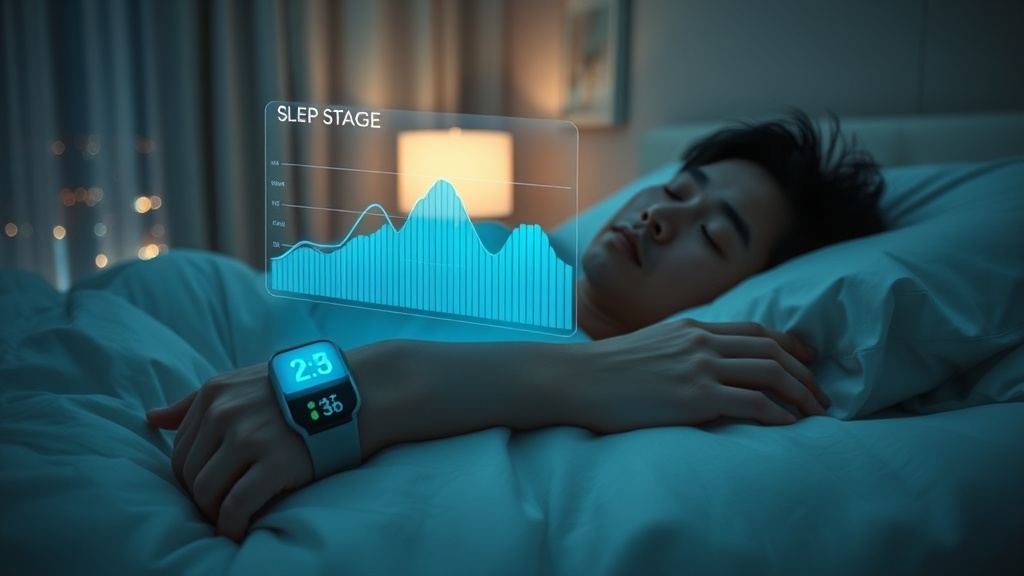
Step 1. Establish your baseline sleep architecture using a validated consumer wearable (Oura, WHOOP, or Apple Watch Ultra). Track for a minimum of 14 consecutive nights. Focus on consistency of sleep efficiency, time in deep sleep, and — if available — HRV trends across the night. These are proxies, not direct arousal measures, but they establish a reference pattern.
Step 2. If you have chronic pain, a diagnosed sleep disorder, or persistent daytime impairment despite "adequate" sleep duration, request a clinical polysomnography with CAP or phasic event analysis. Standard PSG reports often omit microstructural data — you may need to specifically ask for it. This is the only current method that can map A1/A2/A3 subtypes across your night.
Step 3. For those with fibromyalgia or chronic pain conditions showing globally reduced deep sleep metrics, consider evidence-based slow-wave enhancement strategies. Acoustic slow-wave stimulation (pink noise timed to NREM) has shown preliminary efficacy in boosting slow-wave activity, though optimal protocols in pain populations are not yet established.
Step 4. Track your sleep-pain reciprocity systematically. Use a daily pain diary alongside your wearable data. Rate pain intensity (0–10) at wake and before bed for at least 30 days. This longitudinal approach, as advocated by recent perspectives research[3], captures the bidirectional dynamics that single-night studies miss.
Step 5. Prioritize sleep timing consistency over duration. The arousal coupling patterns described by Biabani et al. depend on homeostatic pressure building predictably[1]. Irregular sleep-wake schedules disrupt this buildup. Aim for a consistent bedtime within a 30-minute window.
Step 6. If using HRV as a sleep quality proxy, note the limitation identified in the wearable arousal detection research: HRV features had comparatively lower impact than movement features for predicting cortical arousals[4]. HRV remains useful for autonomic readiness assessment, but don't over-interpret nocturnal HRV dips as direct evidence of arousal events.
Step 7. Reassess every 90 days. Sleep microstructure changes with interventions, seasonal light exposure, and aging. A single baseline is not enough.
Related Video
VERDICT#
6.5/10. This is genuinely interesting descriptive work that reframes how we think about sleep pathology — from stage-level macrostructure to minute-by-minute arousal coupling trajectories. The disorder-specific signatures, particularly fibromyalgia's pan-night A1 deficit, are clinically suggestive. But the sample sizes are small (13–40 per group), the controls were unmatched, no inferential statistics were applied, and the findings are explicitly hypothesis-generating. The companion oscillatory dynamics study adds convergent evidence but shares the same cohort limitations. I appreciate that the authors are transparent about these constraints — that actually increases my confidence in the direction, if not the specific numbers. The wearable detection pipeline isn't mature enough to bring these insights out of the lab yet. Worth watching closely; too early to act on clinically.
Frequently Asked Questions5
References
- 1.Biabani N, Mendonça F, Mutti C. Mapping nocturnal arousal across sleep and pain disorders. Scientific Reports (2026). ↩
- 2.Sicbaldi M, Di Florio P, Palmerini L. Mapping the physiological landscape of body movements during nocturnal sleep and wakefulness and their cardiovascular correlates with a wearable multi-sensor array. Scientific Reports (2025). ↩
- 3.Author(s) not listed. Revealing sleep and pain reciprocity with wearables and machine learning. Communications Medicine (2025). ↩
- 4.Author(s) not listed. Detection of cortical arousals in sleep using multimodal wearable sensors and machine learning. Scientific Reports (2025). ↩
- 5.Biabani N. Disorder-specific alterations of transient oscillatory dynamics during sleep across cortical and subcortical networks. Scientific Reports (2026). ↩
Yuki Shan
Yuki writes with measured precision but genuine intellectual frustration when the data is messy. She uses long, careful sentences for complex mechanisms, then cuts to very short ones for emphasis: 'That's the problem.' She's comfortable saying 'I'm not sure this matters clinically' even when the statistics look impressive. She'll sometimes restart a line of reasoning mid-paragraph: '— actually, I want to rephrase that.' She's suspicious of studies with small sleep cohorts and says so.
View all articles →