Molecular Aging Degradation: Proteostasis, PEP, and Organ Clocks
SNIPPET: Aging degrades the molecular basis of life through a convergent collapse: neuronal protein half-lives double, the protective glycolytic metabolite PEP declines triggering cGAS-STING inflammation, epigenetic entropy accumulates at PRC2 targets, DNA-damaged macrophages drive autoimmunity via autophagy, and organ-specific proteomic clocks now quantify this decay — with brain aging most strongly linked to mortality.
THE PROTOHUMAN PERSPECTIVE#
We talk about aging like it's one thing. It isn't. What these five studies collectively map — published between late 2025 and early 2026 — is a coordinated systems failure at the molecular level, hitting proteostasis, metabolic signaling, epigenetic fidelity, immune tolerance, and senescent cell accumulation simultaneously. This is the degradation layer beneath the wrinkles and the joint pain.
For anyone in the optimization space, the implications are direct. Your neurons are drowning in protein aggregates that microglia can't clear fast enough. Your body's own anti-inflammatory metabolite — PEP — peaks and then abandons you. Your epigenome is losing the instructions that keep cells young. And now we have proteomic clocks precise enough to tell you which organs are failing first, validated across three continents with a cross-cohort correlation of 0.98.
Look, this isn't doom content. It's targeting data. You can't intervene in what you can't measure, and we just got an unprecedented measurement toolkit.
THE SCIENCE#
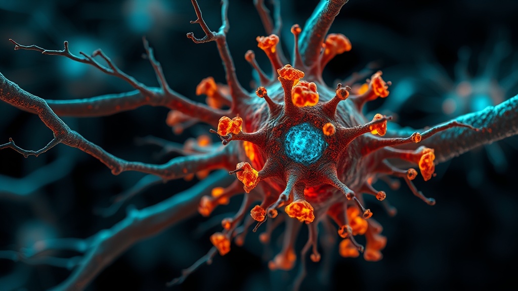
The Neuronal Protein Catastrophe#
The degradation of the proteome with aging isn't gradual — it's a cliff. Researchers publishing in Nature in January 2026 engineered bioorthogonal tagging tools to track nascent neuronal protein turnover in live mice. The core finding: neuronal protein half-life approximately doubles between 4-month-old and 24-month-old mice[1]. That's not a subtle decline. That's the protein recycling machinery running at half speed.
What they call the aged neuronal "aggregome" encompasses 1,726 proteins, and nearly half show reduced degradation with age. Some of these are the usual suspects — proteins already linked to Alzheimer's, Parkinson's, and other neurodegenerative diseases. But many were previously unassociated with neurodegeneration, which opens new target space.
Here's where it gets complicated. These undegraded neuronal proteins accumulate specifically in aged microglia, with 54% also displaying reduced degradation and/or aggregation. Synaptic proteins are heavily enriched in this population, suggesting a cascade: impaired autophagy pathways at the synapse → protein aggregation → microglial engulfment of damaged synapses → synapse loss → cognitive decline[1].
I've been tracking the microglia literature for years. The shift from "microglia as brain janitors" to "microglia as active participants in neurodegeneration" is now backed by hard proteomics. This isn't speculative anymore.
PEP: The Metabolite Your Body Makes — Then Stops Making#
This one caught me off guard. Phosphoenolpyruvate (PEP), a standard glycolytic intermediate that every biochemistry student memorizes, appears to function as an endogenous geroprotector. The team publishing in Nature Aging in March 2026 found a biphasic trajectory: PEP initially accumulates during early aging — apparently as a protective adaptation — then progressively declines[2].
Blocking PEP accumulation exacerbates inflammation and accelerates aging phenotypes. Administering PEP before its natural decline promotes healthy aging in mice.
Wait, let me be more precise here. The mechanism isn't general anti-inflammatory hand-waving. PEP competitively binds to cGAS, the cyclic GMP-AMP synthase that sits at the apex of the cGAS-STING innate immune pathway. The cGAS-STING axis is increasingly recognized as a primary driver of age-related chronic inflammation — it detects cytoplasmic DNA (from mitochondrial damage, nuclear envelope breakdown, etc.) and triggers interferon signaling. PEP essentially jams the sensor[2].
In aged humans, high PEP levels correlated strongly with lower inflammation markers and healthier phenotypic traits. And in a 5×FAD Alzheimer's mouse model, PEP supplementation alleviated neuroinflammation and improved cognitive function[2].
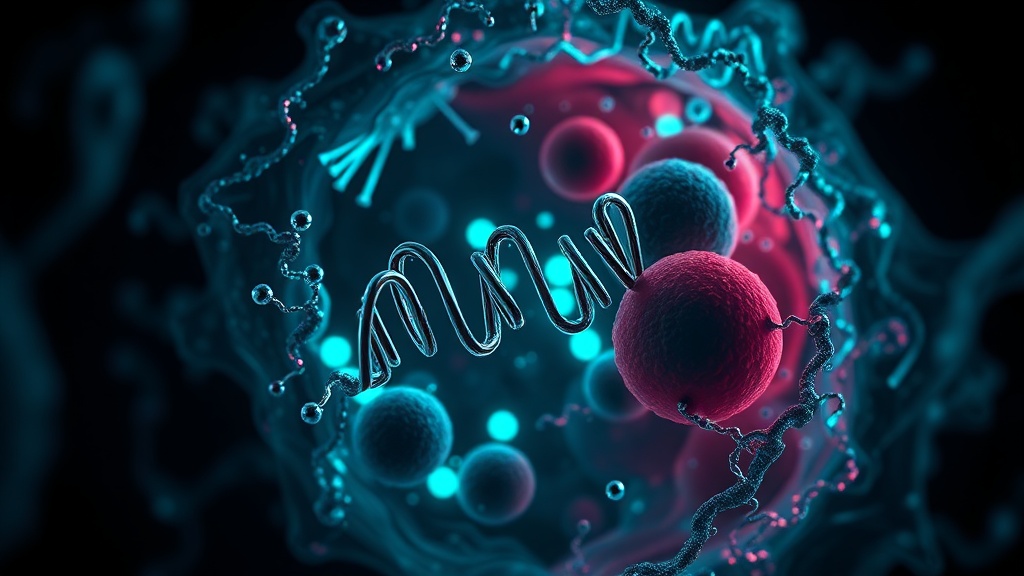
Epigenetic Entropy at PRC2 Targets#
The epigenetic story is equally striking. A February 2026 paper in Molecular Systems Biology used whole-genome bisulfite sequencing to profile DNA methylation changes during aging and partial reprogramming (OSKM factors) in mouse skin[3].
The central finding: aging and rejuvenation converge on the same epigenetic regions — targets of the Polycomb Repressive Complex 2 (PRC2). During aging, PRC2 target regions gain DNA methylation and entropy — meaning the methylation patterns become increasingly disordered. H3K27me3, the histone mark deposited by PRC2, is extensively lost in aged epidermis[3].
Partial reprogramming restores methylation patterns at these exact sites. The boundaries of these disordered regions are defined by large H3K9me2-marked "LOCK" heterochromatin domains — essentially structural borders that contain the epigenetic chaos.
The implication for longevity research is direct: PRC2 activity may be both a readout of aging and a lever for rejuvenation. If you can maintain or restore PRC2 function at these specific genomic regions, you may slow epigenetic drift. This is the kind of mechanistic specificity that moves partial reprogramming from "interesting mouse trick" to "potential therapeutic target."
DNA Damage Drives Autoimmunity Through Macrophage Autophagy#
A January 2026 study in Nature Aging demonstrated that DNA damage in macrophages — accumulated through aging — triggers immune autoreactivity via a specific mechanism: autophagy-mediated nuclear antigen presentation on MHC-II molecules[4].
Using Er1Lyz2/− mice with macrophage-specific DNA repair defects, the team showed that damaged macrophages present nuclear and ribosomal peptides to T cells, driving polyclonal T cell responses and antinuclear autoantibody production. Inhibiting autophagy in these mice suppressed the autoimmune features[4].
The problem with this finding, honestly, is that autophagy is also one of our best anti-aging mechanisms. You need autophagy to clear damaged proteins and organelles. But this study shows that in the context of DNA-damaged macrophages, autophagy becomes the delivery system for self-antigens that trigger autoimmunity. It's a double-edged mechanism, and I'm less convinced we can surgically separate "good autophagy" from "bad autophagy" in aged immune cells without more targeted tools.
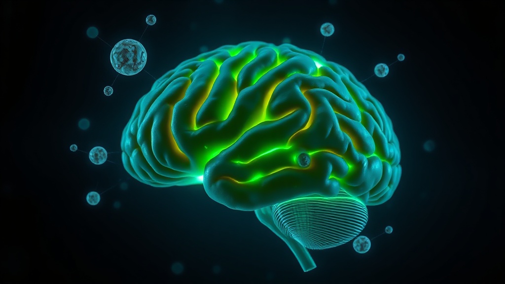
Organ-Specific Proteomic Aging Clocks#
Finally, the measurement revolution. Published in late 2025 in Nature Aging, this study built organismal and ten organ-specific aging clocks using plasma proteomics and machine learning on 43,616 UK Biobank participants, validated in Chinese (n=3,977) and American (n=800) cohorts[5].
Brain aging was most strongly linked to mortality. Accelerated organ aging predicted disease onset, progression, and death beyond clinical and genetic risk factors. A "super-youthful" brain appeared to confer resilience even to APOE4 carriers — the highest genetic risk group for Alzheimer's[5].
The cross-cohort correlation of 0.98 is exceptional for a biological aging metric. This isn't an epigenetic clock that requires tissue biopsy — it's plasma-based, meaning a blood draw could tell you which of your organs is aging fastest.
Key Aging Biomarkers: Magnitude of Change with Age
COMPARISON TABLE#
| Method | Mechanism | Evidence Level | Cost | Accessibility |
|---|---|---|---|---|
| PEP supplementation | cGAS-STING inhibition via competitive cGAS binding | Preclinical (mouse + human correlational) | Unknown (research stage) | Not commercially available |
| Navitoclax (senolytic) | BCL-2 family inhibition | Phase II clinical trials | High (prescription) | Clinical trial only |
| StnIG senotoxin | Pore-forming toxin targeting senescent cell membranes | Preclinical (mouse tumors) | Unknown (research stage) | Not available |
| OSKM partial reprogramming | PRC2 target restoration, epigenetic entropy reversal | Preclinical (mouse) | Extremely high (gene therapy) | Research only |
| Proteomic organ aging clocks | Plasma protein ML-based biological age scoring | Validated in 48,393 humans across 3 cohorts | Moderate (proteomics panel) | Emerging commercial availability |
| Dasatinib + Quercetin (senolytic) | Tyrosine kinase + PI3K inhibition | Small human trials | Low–moderate | Off-label / supplement |
| Autophagy modulation | Clearance of damaged proteins/organelles | Extensive preclinical; mixed human data | Low (fasting, rapamycin) | High (lifestyle) / Moderate (rapamycin Rx) |
THE PROTOCOL#
A practical framework for addressing molecular aging degradation based on current evidence. These steps range from immediately actionable to emerging.
Step 1: Establish Your Baseline with Proteomic Aging Assessment Get a plasma proteomics panel if available through longevity clinics adopting these organ-specific clocks. At minimum, standard inflammatory markers (hs-CRP, IL-6, TNF-alpha) and metabolic panels provide a proxy baseline. Track annually. Knowing which organ system is aging fastest changes your entire intervention strategy[5].
Step 2: Support Proteostasis Through Autophagy — Carefully Time-restricted eating (16:8 minimum, with 2–3 extended 24–36 hour fasts per quarter) activates autophagy pathways critical for clearing aggregated neuronal proteins. However, given the macrophage autophagy–autoimmunity data[4], monitor autoimmune markers (ANA, anti-dsDNA) if you have existing autoimmune tendencies. This is not a blanket "more autophagy = better" situation.
Step 3: Target cGAS-STING Inflammation While PEP supplementation is not yet available, several existing interventions modulate the cGAS-STING axis. Sulforaphane (from broccoli sprouts or standardized extract, 10–20 mg sulforaphane equivalent daily) has shown cGAS-STING suppression in preclinical models. Omega-3 fatty acids (EPA/DHA, 2–3g daily) dampen downstream interferon signaling. These are bridges until PEP-based interventions reach clinical availability.
Step 4: Protect Epigenetic Fidelity at PRC2 Targets Alpha-ketoglutarate (AKG, 300–1000 mg daily) supports TET enzyme function critical for DNA demethylation dynamics. Glycine (3–5g daily) provides methyl group support. Both address epigenetic drift, though neither specifically targets PRC2 restoration — that requires the reprogramming approaches still confined to mouse models[3].

Step 5: Senescent Cell Management The senotoxin StnIG is preclinical[6], but existing senolytic protocols are accessible. Fisetin (500 mg for 2 consecutive days, monthly — based on the Mayo Clinic dosing approach) or quercetin (500 mg) + dasatinib (100 mg) quarterly under physician supervision target accumulated senescent cells that drive SASP-mediated chronic inflammation.
Step 6: Neuroprotective Stack for Synaptic Protein Maintenance Given the microglial accumulation data[1], prioritize interventions that support synaptic health: creatine (5g daily for neuroenergetics), magnesium L-threonate (144 mg elemental Mg, shown to cross the blood-brain barrier), and regular aerobic exercise (150+ minutes/week, which upregulates BDNF and supports microglial homeostatic phenotype).
Related Video
VERDICT#
8.5/10. This cluster of research represents the most mechanistically precise mapping of molecular aging degradation I've seen in a single publication cycle. The PEP–cGAS finding alone could reshape how we think about inflammaging interventions. The proteomic organ clocks are immediately translational. The neuronal aggregome data fills a gap that's been bothering the neurodegeneration field for years. My only reservations: nearly all mechanistic data is still murine, the PEP supplementation pathway in humans is completely uncharted, and the autophagy–autoimmunity paradox complicates one of our most relied-upon longevity strategies. Still — the targeting resolution here is a genuine leap. We're moving from "aging is bad" to "here's exactly which molecular systems fail, in what order, and where to intervene."
Frequently Asked Questions5
References
- 1.Ageing promotes microglial accumulation of slow-degrading synaptic proteins. Nature (2026). ↩
- 2.The glycolytic metabolite phosphoenolpyruvate restricts cGAS-driven inflammation to promote healthy aging. Nature Aging (2026). ↩
- 3.Convergence of aging- and rejuvenation-related epigenetic alterations on PRC2 targets. Molecular Systems Biology (2026). ↩
- 4.DNA damage in macrophages drives immune autoreactivity via nuclear antigen presentation. Nature Aging (2026). ↩
- 5.Organ-specific proteomic aging clocks predict disease and longevity across diverse populations. Nature Aging (2025). ↩
- 6.Senotoxins target senescence via lipid binding specificity, ion imbalance and lipidome remodeling. Nature Aging (2026). ↩
Nael Voss
Nael is data-obsessed and slightly impatient with over-hyped claims. He's tested most of what he covers personally, which means he occasionally contradicts the research when his n=1 doesn't match. His writing moves fast, sometimes too fast — he'll drop a complex mechanism in one sentence and move on. He has a specific verbal tic: 'Look,' when he's about to say something the reader might not want to hear. He's sardonic about supplement marketing but genuinely excited about good mechanistic data.
View all articles →