DBS Precision Neuroimaging Reveals Brain Circuit Map in Parkinson's
SNIPPET: Deep brain stimulation normalizes connectivity in the somatocognitive action network and modulates two distinct neurocircuits — the primary motor and globus pallidus circuits — according to a precision neuroimaging study of 14 Parkinson's patients scanned over one year. Target cortical functional connectivity predicts individual clinical outcomes, opening the door to personalized DBS programming.
Deep Brain Stimulation's Hidden Circuit Map: Precision Neuroimaging Reveals How DBS Rewires the Parkinsonian Brain
THE PROTOHUMAN PERSPECTIVE#
Deep brain stimulation has been a black box for decades. We knew it worked — tremors quieted, rigidity loosened — but the why remained stubbornly opaque. This new research matters because it cracks that box open at the individual level, not the group average. For those of us interested in neural optimization, the implications extend well beyond Parkinson's disease. If we can map precisely how electrical stimulation reshapes functional brain networks in one person across time, we're looking at the foundation for truly personalized neuromodulation — not just for disease, but potentially for cognitive performance, emotional regulation, and the kind of adaptive brain-state management that the next generation of neurotechnology promises. The somatocognitive action network normalization finding is particularly striking: it suggests DBS doesn't just suppress pathological signals but actively restores a network architecture closer to healthy function. That's a qualitatively different story than "turning down the noise."
THE SCIENCE#
What Deep Brain Stimulation Actually Is — And Why We Still Don't Fully Understand It#
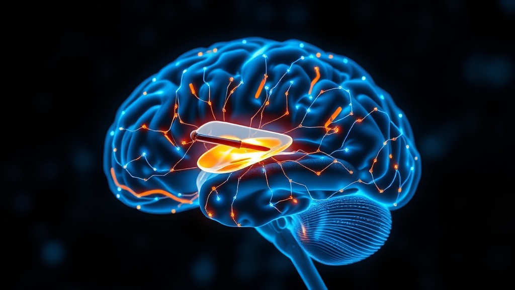
Deep brain stimulation is a neurosurgical intervention that delivers continuous high-frequency electrical pulses through electrodes implanted in targeted brain structures. For Parkinson's disease, the subthalamic nucleus (STN) is the most common target. DBS is one of the most effective therapies for treatment-resistant PD, and it holds promise for depression, Alzheimer's disease, obsessive-compulsive disorder, and epilepsy[1][5].
But here's where it gets complicated. Despite its clinical success, no unifying hypothesis explains how DBS produces its therapeutic effects. Prior imaging studies were limited by small sample sizes, brief scanning sessions, and MRI safety constraints that restricted data quality[1]. The field has been working with blurry snapshots when it needed high-definition film.
The Precision Imaging Breakthrough#
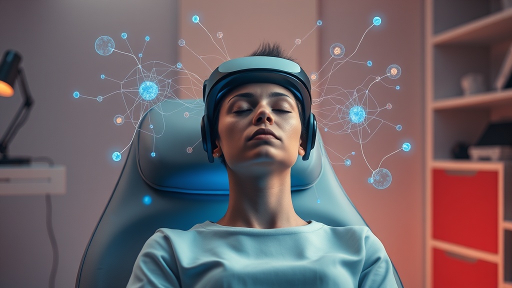
The study published in Nature Neuroscience by researchers using 3-T MRI-compatible DBS represents a genuine methodological leap[1]. Fourteen patients with PD who received subthalamic DBS were scanned extensively across five timepoints spanning one year. The numbers here matter: each patient underwent 11.7 hours of functional MRI under seven different stimulation conditions, plus 2.2 hours of structural MRI and 1.3 hours of diffusion-weighted imaging. Twenty-seven healthy participants served as controls.
I want to sit with that for a moment. 11.7 hours of fMRI per patient. Most DBS-fMRI studies collect perhaps 20–30 minutes. This is an order-of-magnitude difference in data density, and it's what makes individual-level analysis possible rather than relying on group averages that wash out the very individual variation that matters for personalized medicine.
The technical enabler was a new generation of 3-T MRI-compatible DBS leads incorporating helical coils with nonuniform diameters. Previous systems either limited scanning to 1.5-T (lower resolution) or imposed severe restrictions on scan duration for safety reasons[1].
Two Distinct Circuits, One Therapeutic Effect#
The central finding: DBS normalizes functional connectivity in the somatocognitive action network — a large-scale brain network involved in integrating sensory information with motor planning and cognitive control. In PD patients with DBS turned off, this network shows aberrant connectivity patterns compared to healthy controls. With stimulation on, connectivity patterns shift toward the healthy range[1].
But the normalization isn't uniform. DBS evokes differential responses in two distinct neurocircuits: the primary motor circuit and the globus pallidus circuit. This reminds me of something from the attentional blink literature — different context entirely, but the same pattern of a single intervention producing separable downstream effects through distinct pathways.
The primary motor circuit responded more directly to stimulation parameters, while the globus pallidus circuit showed a more complex, potentially modulatory response pattern. What does this actually feel like for patients? The motor circuit effects likely correspond to the immediate tremor reduction patients experience when stimulation begins. The globus pallidus effects may relate to the subtler improvements in gait, posture, and movement initiation that develop over weeks.
Cortical Connectivity Predicts Who Benefits Most#
Perhaps the most clinically actionable finding: target cortical resting-state functional connectivity (RSFC) predicts and tracks motor symptom improvement after STN-DBS[1]. In plain terms, by measuring how the stimulation target connects to cortical regions, clinicians could potentially predict — before or early after surgery — how much benefit a given patient will receive.
This is where the precision imaging approach pays off most directly. Group-level studies can identify general patterns, but they can't tell you whether this specific patient will respond well to this specific electrode placement. The dense individual-level data collected here provides the statistical power to make individual predictions.
The Mechanistic Puzzle: Inhibition, Not Just Modulation#
A complementary study in Nature Neuroscience by a separate research group adds a crucial mechanistic piece[3]. Using fiber photometry and genetically encoded fluorescent sensors in mouse models, they demonstrated that high-frequency DBS of the STN activates afferent axons while simultaneously inhibiting STN neurons. The mechanism? Differential synaptic depression — glutamate release decreases more than GABA release, shifting the excitation/inhibition balance toward net inhibition[3].
I'm less convinced that the mouse model maps perfectly onto the human circuit dynamics described in the imaging study — the species gap is real, and I'd want to see this replicated with human-derived data. But the directionality is consistent: DBS appears to work partly by quieting overactive STN output rather than simply overriding it with electrical noise.
The mouse study also introduces "chemogenetic DBS" — direct chemogenetic inhibition of STN neurons that mimics therapeutic effects without electrical stimulation[3]. This is preclinical data only, but it points toward potentially less invasive future approaches.
The Adaptive Future: Decoding Brain States in Real Time#
A third layer of evidence comes from work on brain signal decoding across 73 neurosurgical patients, published in Nature Biomedical Engineering[4]. This platform integrates machine learning-based brain signal decoding with MRI connectomics across 123 hours of invasively recorded brain data. The connectomics-informed movement decoders generalized across cohorts from the United States, Europe, and China — a critical test of reliability[4].
Horn and Neumann's perspective in Nature Reviews Neurology proposes unifying these threads into what they call "adaptive circuit targeting" — a framework combining adaptive DBS (which adjusts stimulation in real time based on brain state) with connectomic DBS (which identifies optimal circuit targets)[5]. The vision: a system that both knows where to stimulate and when to adjust.
Data Collection per Patient Over 1 Year
COMPARISON TABLE#
| Method | Mechanism | Evidence Level | Cost | Accessibility |
|---|---|---|---|---|
| Precision DBS + fMRI Mapping (this study) | STN stimulation with individual circuit characterization via dense fMRI sampling | Single longitudinal study, 14 PD patients, 27 controls | Very high (research-grade MRI + DBS hardware) | Research settings only |
| Standard DBS Programming | Clinician-guided parameter adjustment based on symptom observation | Decades of clinical use; multiple RCTs | High ($30K–$100K+ surgery; ongoing device costs) | ~200 specialized centers globally |
| Adaptive DBS (aDBS) | Real-time brain signal sensing with automatic stimulation adjustment | Phase II/III trials; emerging clinical data | Very high (next-gen devices required) | Limited to clinical trials |
| Chemogenetic DBS | Targeted chemical inhibition of STN neurons via designer receptors | Preclinical only (mouse models) | Unknown (theoretical) | Not available in humans |
| Levodopa Medication | Dopamine precursor replacement | Gold standard; extensive RCT data | Low–moderate ($50–$300/month) | Widely available globally |
THE PROTOCOL#
For clinicians, researchers, and informed patients navigating the evolving DBS landscape, these findings suggest concrete optimization steps. This is not a DIY protocol — DBS is a surgical intervention requiring specialized medical teams. But understanding the optimization logic matters.
Step 1: Pre-Surgical Connectivity Mapping. Before DBS implantation, obtain high-resolution functional connectivity imaging. The data suggests that baseline cortical RSFC patterns can predict therapeutic outcomes[1]. Request that your surgical team incorporate functional connectivity analysis into the surgical planning pipeline, not just anatomical targeting.
Step 2: Demand Dense Post-Operative Imaging. Standard practice involves limited post-operative imaging primarily to verify electrode placement. Based on this research, advocate for longitudinal functional imaging at multiple timepoints during the first year. Five timepoints (as in this study) across 12 months allows tracking of how brain networks reorganize around the stimulation[1].
Step 3: Individualized Parameter Optimization Using Circuit Response. Rather than relying solely on symptom-based programming (the current standard), push for integration of functional connectivity data into stimulation parameter selection. The finding that different circuits respond differently to stimulation conditions means that a one-size-fits-all approach leaves therapeutic potential on the table.
Step 4: Monitor Both Motor and Somatocognitive Networks. DBS programming typically focuses on motor symptom relief. This study's identification of somatocognitive action network normalization suggests monitoring broader cognitive and sensorimotor integration, not just tremor and rigidity scores[1].

Step 5: Explore Adaptive DBS Eligibility. If you're a candidate for DBS or already have a system implanted, discuss with your neurologist whether next-generation adaptive systems — which sense brain signals and adjust stimulation parameters in real time — may be available through clinical trials[5]. The convergence of connectomic mapping and adaptive stimulation represents the near-term future of the field.
Step 6: Track Subjective Experience Alongside Objective Measures. I think the word "improvement" is doing too much work in most DBS outcome studies. Motor scores matter, but so does the lived experience — initiation of movement, cognitive flexibility, emotional responsiveness. Keep a detailed symptom and experience journal across stimulation conditions to provide your clinical team with data that standard assessments miss.
Related Video
What is the somatocognitive action network and why does DBS normalize it?#
The somatocognitive action network is a large-scale brain network that integrates sensory input with motor planning and cognitive control. In Parkinson's disease, this network shows disrupted connectivity patterns. According to the precision imaging study in Nature Neuroscience, DBS applied to the subthalamic nucleus shifts connectivity in this network toward patterns observed in healthy controls[1]. The normalization may explain why DBS improves not just tremor but also more subtle aspects of movement planning and cognitive-motor integration.
How does differential synaptic depression explain DBS's therapeutic effect?#
High-frequency STN stimulation causes a greater reduction in glutamate release than GABA release at the stimulation site, tipping the excitation/inhibition balance toward net inhibition of STN neurons[3]. This was demonstrated in mouse models using fiber photometry and genetically encoded sensors. Honestly, whether this precise mechanism translates fully to humans is still an open question — the preclinical data is compelling but the species gap shouldn't be minimized.
Why is individual-level brain imaging important for DBS optimization?#
Most prior DBS imaging studies averaged results across patients, which obscures the individual variability that determines whether a specific person benefits from specific stimulation settings. This study collected 11.7 hours of fMRI per patient across seven stimulation conditions, providing enough data to characterize each patient's unique circuit responses[1]. Individual-level cortical connectivity predicted clinical outcomes — something group-averaged data cannot do for a specific patient sitting in front of you.
Who would benefit from adaptive circuit targeting in the future?#
Horn and Neumann's framework suggests that patients with movement disorders, treatment-resistant depression, epilepsy, and potentially Alzheimer's disease could benefit from adaptive circuit targeting[5]. The approach combines real-time brain state decoding with knowledge of which circuits to target for specific symptoms. Early work across 73 patients with brain implants has demonstrated that connectomics-informed decoders can generalize across international cohorts[4], though clinical deployment is still years away.
When will precision DBS imaging become standard clinical practice?#
The honest answer is: not soon enough. The 3-T MRI-compatible DBS technology that enabled this study is relatively new, and the imaging protocols require extensive scanner time that current clinical workflows don't accommodate. I'd estimate 5–10 years before dense functional imaging becomes a routine part of DBS programming at major centers — and longer for community hospitals. But the dataset from this study has been shared publicly, which should accelerate development of clinical tools.
VERDICT#
Score: 8.5/10
This is the most data-rich DBS neuroimaging study I've encountered — the sheer density of individual-level functional data across stimulation conditions and timepoints sets a new standard. The finding that cortical functional connectivity predicts individual clinical outcomes has immediate translational relevance. The complementary mechanistic work on differential synaptic depression adds explanatory depth, even if the mouse-to-human bridge requires caution. I dock points because the sample remains small (14 patients), the technology is currently inaccessible outside research settings, and the somatocognitive network normalization claim — while exciting — needs replication. The public dataset release is a genuinely important contribution that could multiply the study's impact. This is the kind of work that moves DBS from empirical art toward circuit-level engineering.
References
- 1.Author(s) not listed. Circuit response to neuromodulation characterized with simultaneous deep brain stimulation and precision neuroimaging in humans. Nature Neuroscience (2026). ↩
- 3.Author(s) not listed. Differential synaptic depression mediates the therapeutic effect of deep brain stimulation. Nature Neuroscience (2025). ↩
- 4.Author(s) not listed. Invasive neurophysiology and whole brain connectomics for neural decoding in patients with brain implants. Nature Biomedical Engineering (2025). ↩
- 5.Horn A, Neumann W-J. From adaptive deep brain stimulation to adaptive circuit targeting. Nature Reviews Neurology (2025). ↩
- 6.Ye K, Cao Y, Wu Y, Ning C, Wang D, Guo H, Wang Y, Zhang Y, Qiu X, Li N, Wu G, Sun B, Lv X. Therapeutic deep brain stimulation targeting BNST-NAc circuit driven large-scale brain networks in treatment-resistant depression. Translational Psychiatry (2025). ↩
Fen Adler
Fen writes with psychological nuance and a slightly meandering quality that feels human. He'll start pursuing one idea, realize it connects to something else, and follow it briefly before returning: 'This reminds me of something from the attentional blink literature — different context, but the pattern holds.' He's interested in the experience, not just the mechanism, which means he'll occasionally ask: 'What does this actually feel like?' when discussing neurological effects.
View all articles →