ATP Bioenergetics in Depression: Mitochondrial Fatigue Biosignature
SNIPPET: Young adults with major depressive disorder show a distinct ATP biosignature — elevated brain ATP production rates and higher peripheral blood cell ATP levels that correlate directly with fatigue severity. This compensatory mitochondrial overdrive, identified via 7T MRI spectroscopy, masks a reduced reserve capacity, suggesting depression fatigue is a bioenergetic deficit, not psychological weakness.
THE PROTOHUMAN PERSPECTIVE#
Depression fatigue has been dismissed as a motivational problem for decades. That framing is wrong, and we now have the imaging data to prove it.
What Cullen, Tye, Klimes-Dougan et al. published in Translational Psychiatry this month is the first dual-compartment ATP biosignature of fatigue in early-stage major depressive disorder — visible in both brain tissue and circulating blood cells simultaneously[1]. This matters for anyone interested in human performance because it reframes one of the most disabling symptoms in psychiatry as a measurable metabolic event.
If your mitochondria are running harder just to maintain baseline function, you have less reserve for anything else. That's not laziness. That's bioenergetic bankruptcy. And the fact that this signature appears in young adults — mean age 21.8 — tells us the metabolic rot starts early. For the optimization community, the implication is direct: fatigue-associated depression may respond to metabolic interventions that target mitochondrial efficiency, not just serotonin reuptake. The monoamine model isn't dead, but it's increasingly insufficient.
THE SCIENCE#
What ATP Actually Does in the Depressed Brain#
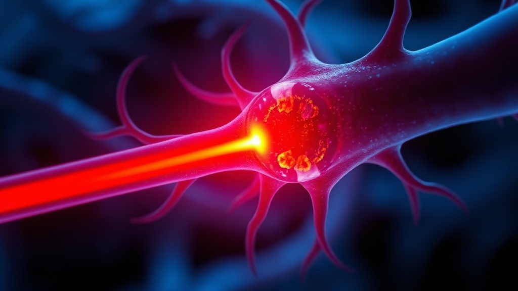
Adenosine triphosphate is the universal energy currency. Every thought, every synaptic transmission, every ion pump cycle burns ATP. The brain consumes roughly 20% of the body's oxygen supply despite constituting only 2% of body mass — a disproportion that makes it exquisitely sensitive to bioenergetic disruptions[3].
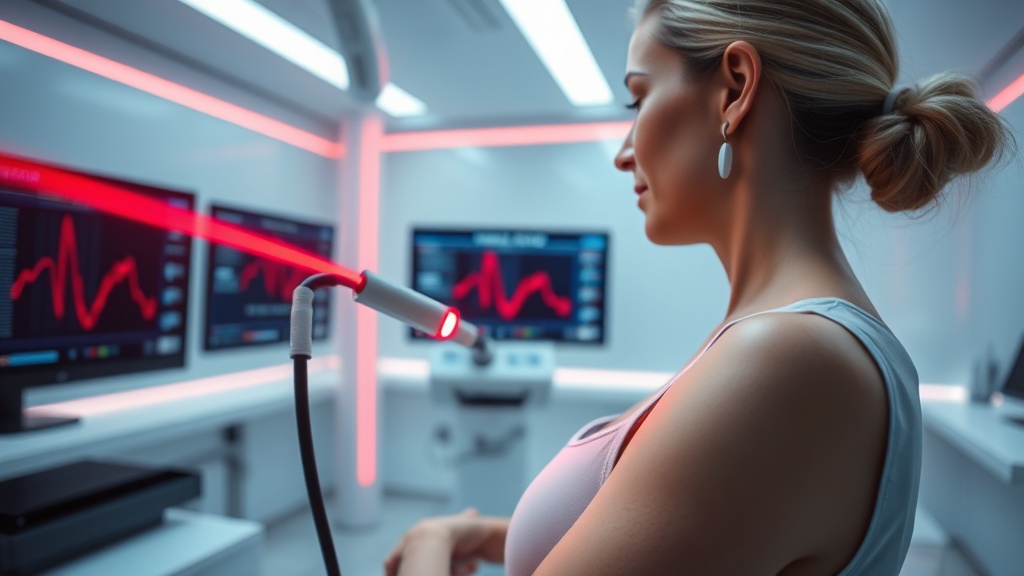
The Cullen et al. study used 31-phosphorus magnetic resonance spectroscopy imaging with magnetization transfer (31P MRSI-MT) at 7 Tesla to measure both ATP concentration and ATP production rate in the visual cortex[1]. This is not your standard clinical MRI. The 7T field strength and the magnetization transfer technique allow quantification of the forward rate of the creatine kinase reaction — essentially measuring how fast the mitochondrial machinery churns out ATP in real time.
Nine MDD participants. Nine healthy controls. Small sample. I'll address that.
The Paradox: More Production, Less Reserve#
Here's where the data gets interesting and uncomfortable at the same time.
The MDD group showed higher ATP production rates in the visual cortex than healthy controls. Not lower. Higher. And this elevated production rate correlated positively with Fatigue Severity Scale (FSS) scores — meaning the more fatigued a participant felt, the faster their brain was synthesizing ATP[1].
The parallel finding in peripheral blood mononuclear cells (PBMCs) mirrored this pattern. Resting ATP concentrations were higher in the MDD group's PBMCs than in controls, and again correlated with fatigue severity. But here's the catch: after mitochondrial uncoupling — a stress test that forces the electron transport chain to maximum output — PBMCs from the MDD group had a lower capacity for ATP production than healthy controls[1].
This is the compensatory mechanism the authors describe. At baseline, the depressed brain and immune cells are working harder to maintain normal ATP levels. They're revving the engine in third gear because fifth gear is broken. The reserve capacity — the headroom that lets you handle cognitive load, physical exertion, emotional stress — is depleted.
The Blood Mirror#
The PBMC findings deserve separate attention because blood is accessible. Brain spectroscopy at 7 Tesla is a research tool — you're not getting this at your local imaging center. But a blood draw? That's scalable.
The fact that peripheral blood cells reflect the same ATP dysregulation seen in the brain suggests a systemic bioenergetic phenotype in early MDD. This aligns with a separate meta-analysis by Chen et al. published in Translational Psychiatry in February 2026, which found elevated circulating cell-free mitochondrial DNA (ccf-mtDNA) significantly associated with MDD (p = 0.013), with particularly strong associations in unmedicated patients (p = 4.99 × 10⁻⁶)[2]. Mitochondria under stress leak their DNA into the bloodstream. The ATP data from Cullen et al. and the ccf-mtDNA data from the meta-analysis are telling the same story from different angles: mitochondrial dysfunction in depression is systemic, measurable, and present before chronic-stage disease.
A third study from Wen et al. in Nature Mental Health adds further weight, using network control theory to demonstrate that MDD brains exhibit energy inefficiency — elevated energy costs with reduced control stability — particularly in the left dorsolateral prefrontal cortex and insula[4]. The serotonin 5-HT2A receptor and astrocyte dysfunction were linked to these energy deficits.
The CD8+ T-Cell Connection#
Huang et al.'s work on mitochondrial mass in CD8+ T-cell subsets in depressed patients found that mitochondrial mass increased significantly in several CD8+ subsets after treatment, while the percentage of cells with low mitochondrial membrane potential decreased — but only in patients hospitalized for 21 days or fewer[5]. Longer hospitalization showed no such recovery. This suggests there's a window where mitochondrial function in immune cells can rebound with treatment, and that window may close.
Let me push back on something, though. The Cullen et al. study had 18 usable imaging participants and 24 for the PBMC analysis. That's a pilot. The correlations are biologically plausible and the dual-compartment design is clever, but I'd want to see this replicated with n > 50 before anyone redesigns clinical protocols around it. The effect could be real and still not survive a larger, more diverse sample.
ATP Biosignature in MDD vs. Healthy Controls
Bioinformatics Confirmation#
Chen Z. et al.'s bioinformatics analysis identified four mitochondria-associated biomarkers for MDD — SLC25A5, ALDH2, CPT1C, and IMMT — with AUC values exceeding 0.7 in two independent datasets[6]. SLC25A5 encodes the adenine nucleotide translocator, directly involved in ATP/ADP exchange across the inner mitochondrial membrane. Its dysregulation in MDD isn't surprising given the ATP data, but it provides a genetic hook for the metabolic phenotype Cullen et al. observed at the functional level.
COMPARISON TABLE#
| Method | Mechanism | Evidence Level | Cost | Accessibility |
|---|---|---|---|---|
| 31P MRSI-MT at 7T (Cullen et al.) | Measures brain ATP production rate via creatine kinase flux | Pilot study, n=18 | ~$2,000–4,000/scan | Research-only; <100 sites globally |
| PBMC ATP analysis (Cullen et al.) | Measures resting and stressed ATP in blood immune cells | Pilot study, n=24 | ~$200–500 | Specialized labs; potentially scalable |
| ccf-mtDNA blood test (Chen Y. et al. meta-analysis) | Quantifies leaked mitochondrial DNA as stress marker | Meta-analysis, 13 studies, n=1,370 | ~$100–300 | Clinical labs; most scalable |
| MitoTracker flow cytometry (Huang et al.) | Measures mitochondrial mass and membrane potential in T cells | Observational, n=40 | ~$150–400 | Hospital labs with flow cytometry |
| Bioinformatics gene panels (Chen Z. et al.) | Identifies MRG/ARG expression biomarkers | Computational validation | ~$300–800 (expression panels) | Research and specialized diagnostic labs |
THE PROTOCOL#
Based on current evidence — and I want to be clear this is informed by early-stage data, not multiple confirmed RCTs — here's a practical framework for addressing mitochondrial bioenergetic deficits associated with depression fatigue.
Step 1: Get a baseline assessment. Request inflammatory markers (hs-CRP, IL-6) and a metabolic panel. If accessible, ask about PBMC mitochondrial function testing — some integrative medicine clinics now offer mitochondrial stress tests via the Seahorse XF platform or equivalent. Document your fatigue using the Fatigue Severity Scale (FSS), a validated 9-item questionnaire.
Step 2: Support NAD+ synthesis pathways. NAD+ is the primary electron carrier in oxidative phosphorylation. Nicotinamide riboside (NR) at 300 mg/day or nicotinamide mononucleotide (NMN) at 250–500 mg/day, taken in the morning, may support mitochondrial NAD+ pools. Optimal dosing in humans for depression-related fatigue is not yet established — this is based on general mitochondrial support literature, not MDD-specific trials.
Step 3: Address mitochondrial cofactors. Coenzyme Q10 (200–400 mg/day with a fat-containing meal), magnesium glycinate (300–400 mg/day, evening), and B-complex vitamins — particularly B2 (riboflavin, a Complex I/II cofactor) and B3 — form the substrate foundation for electron transport chain function.
Step 4: Implement controlled aerobic exercise. This is the single intervention with the strongest evidence for improving mitochondrial biogenesis via the PGC-1α pathway. Start with 20–30 minutes of zone 2 cardio (nasal breathing pace), 3–4 times per week. Depression makes this difficult. Start absurdly small if needed — 10 minutes of walking counts.

Step 5: Prioritize sleep architecture. Mitochondrial autophagy (mitophagy) — the clearance of damaged mitochondria — is upregulated during deep sleep. Target 7–9 hours with consistent sleep-wake times. Consider tracking HRV as a proxy for autonomic and mitochondrial recovery; a declining HRV trend may indicate inadequate mitochondrial repair.
Step 6: Monitor and reassess at 8 weeks. Repeat FSS scoring. If accessible, repeat inflammatory markers. Adjust protocol based on response. If fatigue persists despite metabolic support, this is not a failure of willpower — it may indicate the reserve capacity deficit is deeper than lifestyle interventions can address, and pharmacological options should be discussed with a clinician.
Step 7: Consider red/near-infrared light exposure. Photobiomodulation at 630–670 nm and 810–850 nm has preclinical evidence for enhancing cytochrome c oxidase activity (Complex IV), the terminal enzyme in the electron transport chain. Wavelength matters. Irradiance matters. Time matters. Most consumer devices get at least one of these wrong. Target 10–20 mW/cm² at the tissue surface, 10–20 minutes per session, applied to the forehead or neck for potential transcranial and vagal effects.
Related Video
What is the ATP biosignature of depression fatigue?#
It's a pattern first identified by Cullen, Tye, Klimes-Dougan et al. in 2026 where young adults with MDD show elevated ATP production rates in the brain and elevated resting ATP in blood cells — but reduced ATP reserve capacity under stress. The elevated baseline production appears to be a compensatory response to inefficient mitochondria, not a sign of healthy energy metabolism.
Why does higher ATP production correlate with worse fatigue?#
Because the higher production rate reflects the system working harder to maintain normal output, not producing surplus energy. Think of it as a car engine running at 5,000 RPM to maintain 60 mph — the engine is under stress even though the speed looks normal. When demand increases, there's no headroom left, and fatigue is the subjective experience of that bioenergetic ceiling.
How could a blood test detect depression fatigue?#
PBMCs — the immune cells in your blood — showed the same ATP dysregulation as brain tissue in this study. The ccf-mtDNA meta-analysis by Chen et al. (n=1,370) also found elevated mitochondrial DNA fragments in blood of unmedicated MDD patients[2]. A scalable blood-based test combining these markers could potentially identify bioenergetic depression subtypes, though clinical validation is still needed.
Who would benefit most from mitochondrial-targeted depression treatment?#
Based on current data, young adults with early-stage MDD who present with fatigue as a primary symptom and who have not yet been treated with antidepressants may show the clearest bioenergetic abnormalities. The ccf-mtDNA meta-analysis found the strongest associations in unmedicated patients (p = 4.99 × 10⁻⁶), while medicated patients showed no significant association[2]. This suggests antidepressants may partially normalize mitochondrial stress markers.
When will mitochondrial biomarkers be available in clinical practice?#
Honestly, we don't know yet. The 7T spectroscopy technique is purely research-grade. Blood-based markers like ccf-mtDNA and PBMC ATP analysis are technically feasible now but lack standardized clinical protocols. I'd estimate 3–5 years before any of these reach diagnostic use, assuming replication studies confirm the findings and someone builds the clinical infrastructure.
VERDICT#
7.5/10. The dual-compartment ATP biosignature is a genuinely novel finding — I haven't seen another study capture compensatory mitochondrial overdrive in both brain and blood simultaneously in early MDD. The biological logic is tight: elevated baseline, depleted reserve. The convergence with the ccf-mtDNA meta-analysis and the Wen et al. energy inefficiency data from Nature Mental Health builds a coherent picture. But the sample size is a problem I can't ignore. Eighteen imaging participants isn't enough to establish a biomarker — it's enough to justify the next study. The PBMC data is more actionable because it's scalable, but even 24 participants leaves too much room for noise. I'm cautiously optimistic and genuinely interested in the replication. The compensatory mechanism hypothesis, if confirmed, would fundamentally change how we think about depression fatigue. That's worth watching.
References
- 1.Cullen KR, Tye SJ, Klimes-Dougan B. ATP bioenergetics and fatigue in young adults with and without major depression. Translational Psychiatry (2026). ↩
- 2.Author(s) not listed. Blood circulating cell-free mitochondrial DNA as a potential biomarker for major depressive disorder: a meta-analysis. Translational Psychiatry (2026). ↩
- 3.Erecińska M, Silver IA. ATP and brain function. Journal of Cerebral Blood Flow & Metabolism (1989). ↩
- 4.Wen Y. Energy inefficiency underpinning brain state dysregulation in individuals with major depressive disorder. Nature Mental Health (2026). ↩
- 5.Huang Y, Chen Z, Xu Y, Ren B. Mitochondrial mass and low mitochondrial membrane potential percentage of CD8+ T cell subsets are implicated with therapeutic effect in depressive disorder. Frontiers in Psychiatry (2025). ↩
- 6.Chen Z, Tang X, Gu C, Zou S. Investigating biomarkers of mitochondrial and aging-related genes in major depressive disorder through bioinformatics analysis. Frontiers in Psychiatry (2025). ↩
Sova Reld
Sova writes with focused intensity and low tolerance for vague claims. She came to photobiomodulation through personal experimentation and is irritated by both true believers and reflexive skeptics. Her writing has edge: 'The wellness market has done more damage to this field than the skeptics ever could.' She's extremely precise about parameters — wavelength, irradiance, duration — and will tell you when a study used inadequate dosing without apology.
View all articles →